- Main
- Useful Information
- Ultrasound in the early stages of pregnancy
Ultrasound in the early stages of pregnancy
Ultrasound in the early stages of pregnancy is recommended to every woman, as it is the easiest and safest method. The study helps to confirm the fact of pregnancy, can show whether the embryo is developing correctly, allows to see and eliminate problems in time, if any.
Ultrasound waves have proven safety and cannot harm the baby.
The purpose of ultrasound in the early stages of pregnancy
At the initial stage of pregnancy, ultrasound diagnostics has 2 main indications:
- confirmation of a successfully developing pregnancy. It is carried out in 4-6 weeks, most often it happens during the first visit of a woman to a gynecologist when menstruation is delayed
- scheduled first pregnancy screening. It is prescribed for 11-13 weeks in order to assess the pace of development, exclude ultrasound signs of genetic abnormalities and congenital pathologies.
It is recommended to do an ultrasound in the early stages of pregnancy at the 1st visit with a delay of 7 days or more (to rule out an ectopic pregnancy).
If the results of scheduled examinations do not reveal abnormalities, correction of pregnancy management is not required. Indications for an unscheduled ultrasound during pregnancy can be pain in the lower abdomen, vaginal bleeding, deviations according to the results of laboratory tests.
Features of ultrasound in the early stages
Conducting ultrasound in the early stages of pregnancy gives the doctor the necessary information about the development of the embryo:
- the location of the fetal egg in the uterus (normal) or in the fallopian tubes (ectopic pregnancy that requires timely medical assistance);
- elucidation of the number of viable embryos and features of attachment to the uterine wall;
- listening to the baby's heartbeat, which becomes possible as early as the 4th-5th week of pregnancy;
- determining the proportionality of embryo development, measuring the linear parameters of the body and the thickness of the cervical fold.
At the Giorno Medical Center, pregnant women have access to an ultrasound service using a modern machine that provides the most accurate information in the early stages. The clinic has a friendly atmosphere, the staff cares about the comfort of expectant mothers and the quality of the examination performed.
How ultrasound is performed in the early stages of pregnancy
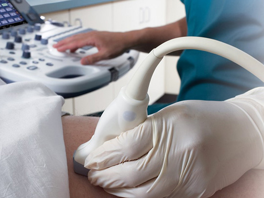
Depending on the indications for ultrasound diagnostics in pregnant women, the transabdominal method is used, in which the sensor is applied to the front surface of the abdominal wall, and the transvaginal method is inserted into the vagina. What method to choose in each specific case, the doctor decides after interviewing and examining the pregnant woman.
To perform a transabdominal ultrasound, a specialist applies a gel conductor to the skin of the abdomen and attaches a sensor. By changing the angle of inclination and the scanning plane, the doctor gets a clear picture of the uterus, the contours of the fetal egg and the embryo on the monitor screen.
To perform a transvaginal ultrasound in the early stages of pregnancy, the doctor puts a condom on the sensor, lubricates it with gel and gently inserts it into the vagina. The tip is located shallowly and causes unpleasant sensations in the woman. The specialist slightly deviates the sensor to the sides to examine the uterus with appendages and other structures, to see the fetal egg and its location. The study takes 10-15 minutes.
Contraindications to ultrasound in the early stages of pregnancy
Ultrasound is contraindicated:
- at the threat of spontaneous abortion
- in the presence of severe inflammatory processes in the vagina
- in cases of psychological discomfort from the manipulation of a pregnant woman.
How to prepare for ultrasound?
Before going to the doctor, it is necessary to perform standard genital hygiene, no other preparation is required.
Can ultrasound harm a child?
The safety of the study is confirmed by authoritative organizations: ultrasound is recommended by the World Health Organization as one of the key screening methods for pregnancy pathologies and fetal development anomalies, which increases the likelihood of a healthy baby and helps avoid many complications.
The results
At the end of the ultrasound examination, the doctor attaches the printed pictures. Immediately after the examination, the doctor draws up a medical document, which indicates the term and features of the pregnancy, the number of embryos, the state of the woman's reproductive organs, etc.
Screening ultrasound of the first trimester also allows you to evaluate the function of closing the cervix to exclude isthmic-cervical insufficiency.
Doppler imaging of the uterine arteries, performed in the first trimester, helps to determine the risk of preeclampsia - a complication of pregnancy in the second trimester, which is characterized by an increase in blood pressure and the appearance of protein in the urine above acceptable standards.


