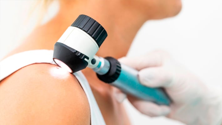
Dermatoscopy of the skin. When it should be done
Dermatoscopy of the skin is a highly informative instrumental method of non-invasive examination of the skin. The study allows you to evaluate any formations on the skin and determine their nature (benign or malignant). One of the main advantages is the early detection of skin cancer, in particular, melanoma. The method allows to increase the timely detection of the disease by 20%.
The purpose of dermatoscopy of the skin
Despite the multitude of indications, dermatoscopy of moles is most often performed in order to determine their malignancy. A specialist can prescribe the procedure for the following changes in pigmented nevi:
- uneven growth on the surface of the skin;
- lack of clear borders with healthy tissues;
- heterogeneous coloring (especially dangerous black-blue shades);
- large size (diameter over 6 mm);
- constant change in the shape, color and size of education;
- itching, burning and pain in the area of the pigment spot;
- the presence of papillomatous growths;
- bleeding and ulcers of any size;
- lack of hair growth in the area of the changed skin area.
The clinical manifestations described above may be the first signs of malignant transformation of moles.
When they are detected, it is recommended to urgently consult a dermatologist or oncologist, who will conduct a dermatoscopy of the nevus and draw up a plan for further actions. Also, as a method of diagnosis, it is recommended for all persons with light skin, blue eyes and swallow once a year. It is this group of people who are at risk of developing melanoma.
Indications for dermatoscopy of the skin
Dermatoscopic examination is used to diagnose various skin lesions. The main indications include:
- differential diagnosis between benign and malignant neoplasms;
- early detection of melanoma;
- confirmation of parasitic skin pathologies (acarodermatitis or scabies, pediculosis);
- identification and differentiation of bacterial, viral and fungal skin infections;
- assessment of the condition of the nail plates;
- search for the cause of alopecia.
The method is recommended to be used in the case of regular exposure to risk factors that provoke the malignant transformation of mole cells:
- frequent exposure to the sun;
- regular visits to cosmetology centers;
- any hormonal disorders in the body (including pregnancy, menopause and teenage periods);
- mechanical damage to pigment nevi.
Algorithm of the doctor's actions during the examination:
- Search for formations on the skin and their visual assessment. In some cases, a detailed examination (with a clear clinical picture) is not necessary.
- Application of specialized gels. They allow you to increase the transparency of fabrics and eliminate glare (their absence is important when taking pictures).
- Full illumination of the lesion. Some models of dermatoscopes are already equipped with a lighting device. To increase the value of dermatoscopy of neoplasms, pressing of the focus with a glass can be performed (in the absence of secretions, ulcers and bleeding).
- Careful study of pathological inclusions and execution of a series of images.
- Computer evaluation of changes in skin thickness.
- Making a diagnosis and developing further tactics of diagnostic and treatment measures.


